US$700.00-
Recombinant mouse IgGI, 50μg
Product Code: RCA-001
* This product can be purchased directly from us
・Leaflet  | ・References | ・How to Order
Overview:
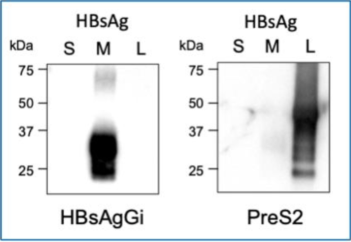
HBV envelope protein is composed of three types of surface antigens, S-, M-, and L-HBs, which are produced from an HBsAg gene containing PreS1, PreS2 and S-domains1. HBsAgs are heavily glycosylated with N-glycan and O-glycan2,3. Whole glycan structural analyses revealed that PreS2 domain on M-HBs, but not on L-HBs, contains highly conserved O-glycosylated site in genotype C (gC)4,5.
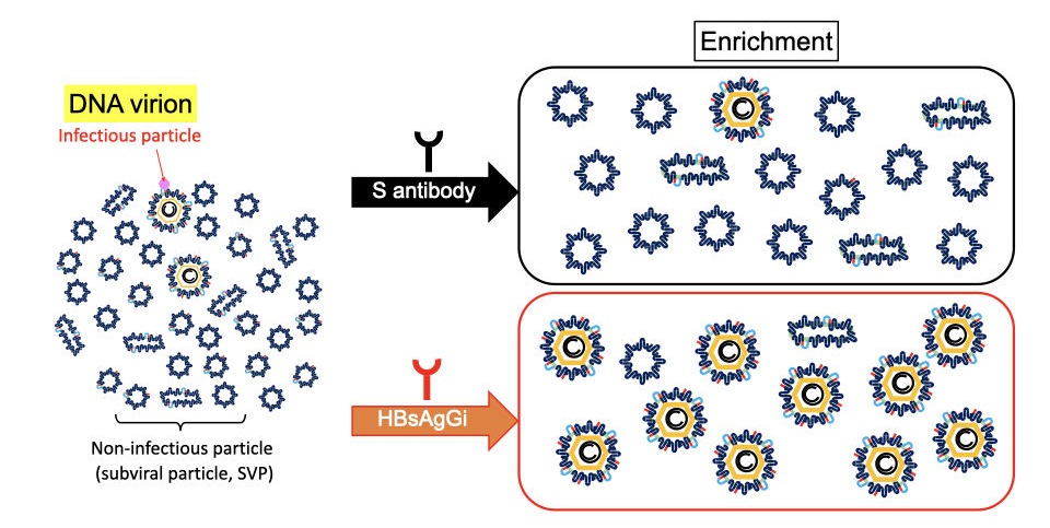
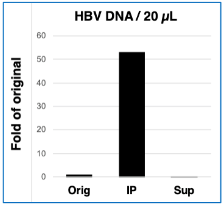
A recombinant monoclonal antibody against HBsAg glycan isomer (HBsAgGi) was generated by using O-glycosylated PreS2 peptide. Compared to traditional HBsAg-testing which recognizes all viral particles, HBsAgGi specifically recognizes infectious HBV particles (DNA virion).
Isotype: mouse IgG1
Light chain: kappa
Amount: 50 μg
Storage:Store at 4°C for short term. Upon delivery store at -20°C for long term and avoid freeze / thaw cycle.
Applications:
1. Western
2. Immunoprecipitation/Separation of HBV particles
3. ELISA
4. HBV infection inhibition assay
5. Immunohistochemistry
HBV virion contains envelope protein (HBsAg), HBV core and HBV DNA. HBsAg gene consists of PreS1 (blue), PreS2 (red), and S (black) -domains. About half of S-HBsAgs is modulated by N-glycan and the rest is non-glycosylated. PreS2 domain in M-HBsAg is highly O-glycosylated, but not in L-HBsAg. S-HBsAg is the most abundant and forms non-infectious particles. Infectious HBV particles consist of all HBsAgs and contains HBV DNA. HBsAgGi recognizes O-glycosylated PreS2 domain in M-HBsAg of genotype C.
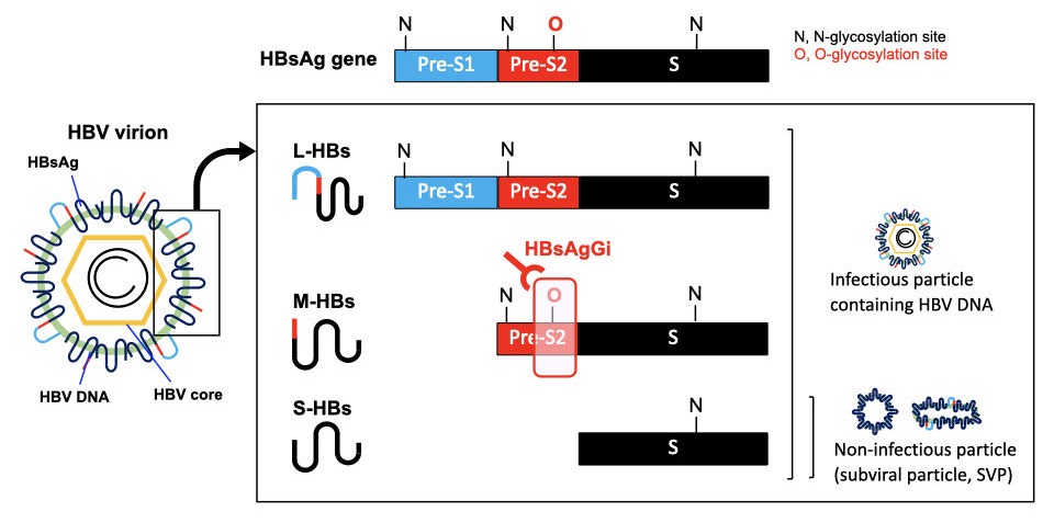
In HBV patients’ serum, non-infectious particles are much dominant than infectious particles containing HBV DNA. Anti-S antibody may recognize all HBV particles including non-infectious particles, However, HBsAgGi specifically recognizes HBV particles containing M-HBsAg, resulting in efficient recognition of infectious HBV particles. HBsAgGi measuring will classify HBV patients differently from the S-antibody measuring.
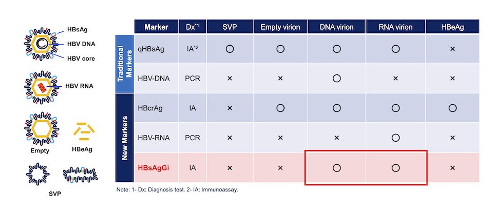
Comparison of HBV markers. Measuring qHBsAg and HBV DNA have been used, while HBcrAg and HBV RNA are new markers to diagnose patient condition. Target HBV particles or molecules are indicated in the table. Circle (〇) indicates the target particle and molecules, and X indicates that the marker cannot recognize or not recommended.
HBsAgGi detects M-HBsAg-containing particles such as DNA or RNA virion.
Examples:
References:
1. Schadler and Hildt (2009) Viruses 1:185-209.
2. Schmitt et al. (2004) J Gen Virol 85:2045-2053.
3. Dobrica et al. (2020) Cells 9:1404.
4. Wagatsuma et al. (2018) Anal Chem 90:10196-10203.
5. Angata et al. (2021) Biochim Biophys Acta Gen Subj. 1866:130020
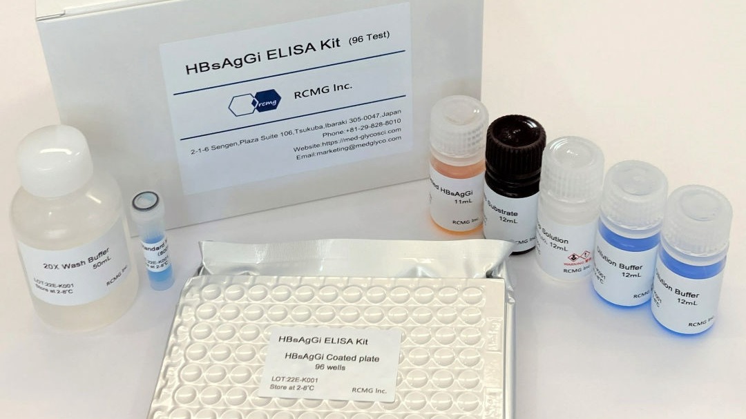
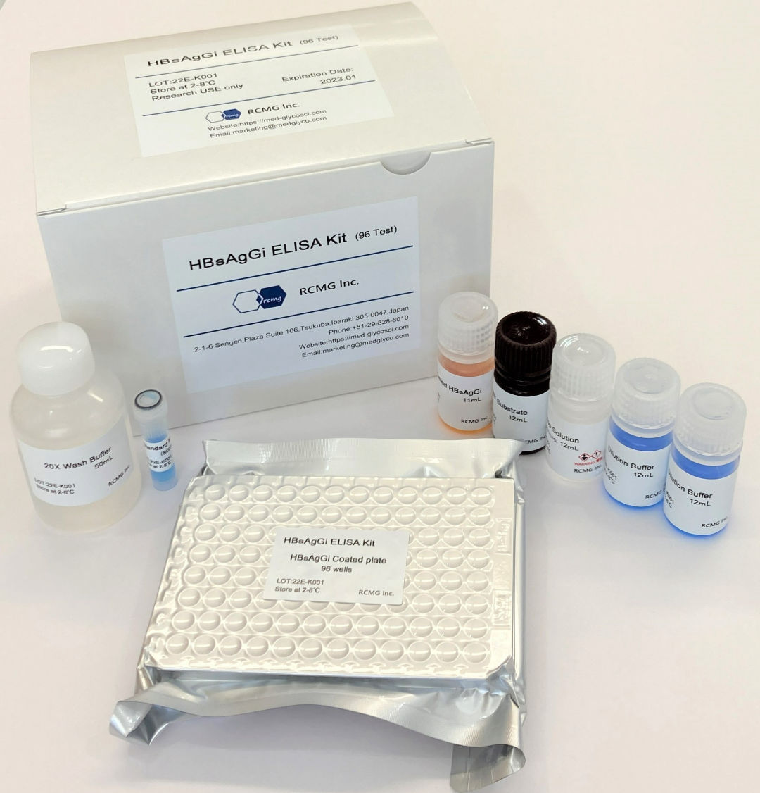
 HBsAgGi ELISA Kit:
 US$1,000.00-
96 Test
Product Code: RCEK-001
* This product can be purchased directly from us
・Leaflet | ・Safety data sheet / User guide |
・References | ・How to Order
Overview:
A recombinant monoclonal antibody against HBsAg glycan isomer (HBsAgGi) is specific to O-glycosylated Pre-S2 on M-HBs, which are predominantly present in infectious HBV particles. Compared to traditional HBsAg-testing which recognizes all viral particles, HBV particles captured by HBsAgGi differ from those captured by S-HBsAg antibody.
By using HBsAgGi, we established for the first time a new ELISA system to measure HBV DNA-containing viral particles. Sera of HBV patient mainly contains subviral particles over the HBV DNA particles. The ratio of infectious particles vs non-infectious particles differs in each patient. Thus, measuring infectious HBV particles would be useful to analyze pathological conditions of patients.
Materials included in the kit
HBsAgGi antibody coated plate:96 well (8-well strip x 12)
Standard M-HBsAg
Dilution Buffer
20X Wash Buffer
HRP-labeled HBsAgGi antibody
TMB Substrate
Stop Solution
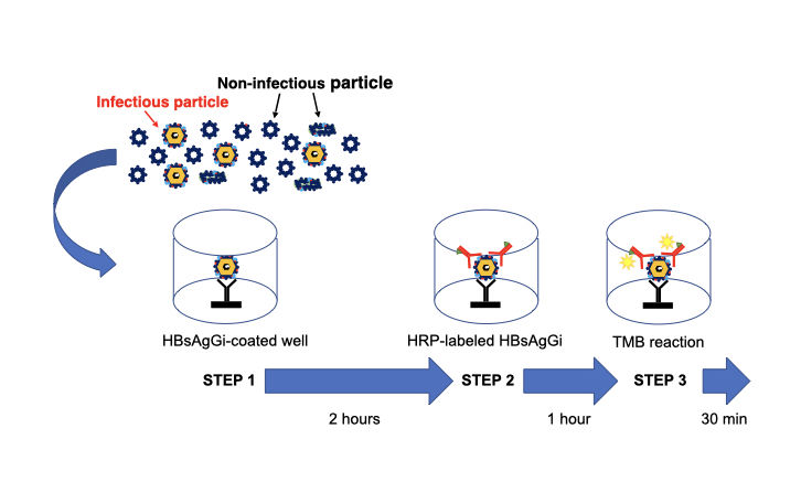
HBV DNA particles can be mainly captured by HBsAgGi antibody (STEP 1). After washing, the HBV particles captured on the ELISA microwells will be further reacted with the HRP-labeled HBsAgGi antibody, (STEP 2). After further washing, HRP substrate (TMB substrate) is added into the well. After the incubation, the reaction is stopped and the absorbance at 450 nm is measured by a plate reader (STEP 3).
Examples:
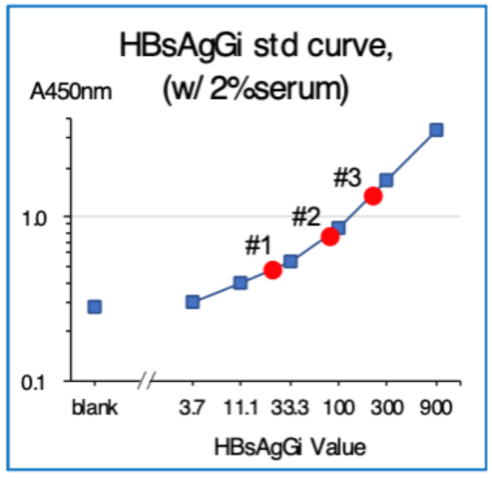
Standard curve
Standard curve was obtained by using Standard M-HBsAg included in the kit.
Standard M-HBsAgs (6.25 – 400 ng/mL) were obtained by serial dilution of M-HBsAg (800 ng/mL).
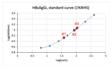
| Sample | qHBsAg (IU/mL) | HBsAgGi (µg/mL) | CV (%) |
|---|---|---|---|
| Serum #1 | 2100 | 4.5 | 2.52 |
| Serum #2 | 8200 | 8.92 | 2.65 |
| Serum #3 | 2200 | 11.04 | 1.82 |
References:
1. Schadler and Hildt (2009) Viruses 1:185-209.
2. Schmitt et al. (2004) J Gen Virol 85:2045-2053.
3. Dobrica et al. (2020) Cells 9:1404.
4. Wagatsuma et al. (2018) Anal Chem 90:10196-10203.
5. Angata et al. (2021) Biochim Biophys Acta Gen Subj. 1866:130020
2-1-6 Sengen, Plaza Suite 106, Tsukuba, Ibaraki 305-0047
TEL:+81-29-828-8010
marketing@rcmg-glyco.com
Service
Home
About Us
Products & Services
• Research Products
• Contract Services
Projects
News
Access
Contact
Japanese Website
© Copyright 2022 RCMG Inc.
Contact Us